| C-Trp
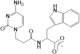
Cytosine-1-yl-(2-carboxyethyl)-L-tryptophan hexahydrates 
M.Doi, M.Tarui, M.Ogata, A.Asano & T.Ishida (1998) Acta Crystallogr. Sect.C, 54, 1941-1943. This study was supported by the Grant-in-Aid for Scientific Research 07672427 from the Ministry of Education, Science, Sports and Culture. |
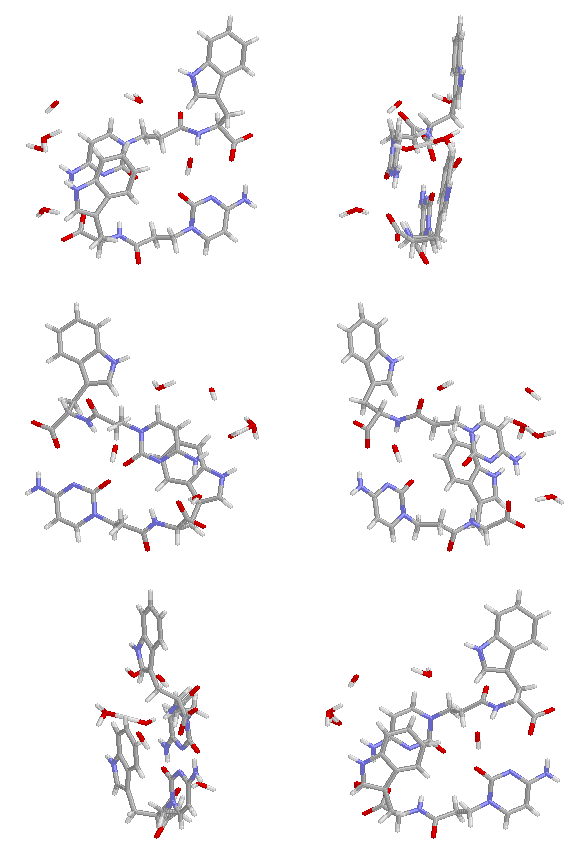
 This structer was produced by RasMol, and stucked by GifBuilder (Yves Piguet, 1997) |
The title compound C-Trp was designed to investigate the cooperative interactions between the nucleic acid and polypeptide, and the ability of H-bond and pai-pai stacking between the nucleic base and functional group of peptide was expected. The structure property is similar to that of peptide nucleic acid (PNA). It is known that the indole ring hybridized with nucleic base has the high affinity against the N7-quarternized guanine.
The hybrid dipeptide containing cytosine base and L-tryptophan was crystallized as a hexahydrates form, and the solvent occupied 13% of asymmetric weight. Two independent molecules were distinguished by the direction of indole ring. All polar atoms of the title compound participated in H-bond formation, and the tight network was built by combination with solvent-mediated H-bonds. The pai-pai electron interaction was observed between the indole and nucleic base, which was facilitated by H-bonds.
Related Papers:
Ishida, T., Iyo, H., Ueda, H., Doi, M., & Inoue, M. (1990). J. Chem. Soc., Chem. Commun. 217-218.
Ishida, T., Tarui, M., In, Y., Ogiyama, M., Doi, M., & Inoue, M. (1993). FEBS Lett. 333, 214-216
Kamiichi, K., Danshita, M., Minamino, N., Doi, M., Ishida, T., & Inoue, M. (1986). FEBS Lett. 195, 57-60.
Kamiichi, K., Doi, M., Nabae, M., Ishida, T., & Inoue M. (1987). J. Chem. Soc., Perkin Trans. II 1739-1745.
Related compounds:
C-Ile (cytosinyl-L-isoleuicine)
C-Ala (cytosinyl-L-alanine)
C-Tyr (cytosinyl-L-tyrosine)
C-Phe (cytosinyl-L-phenylalanine)
C-Asp (cytosinyl-L-asparadic acid)
C-Ser (cytosinyl-L-serine)
X-Ray Data Summary
| formula | 2(C18H19N5O4).6H2O | ||
| weight | 383.37 | ||
| symmetry | triclinic | ||
| space group | P1 | ||
| Cell | Crystal | ||
| a | 9.2046(7) Ang. | description | plate/td> |
| b | 14.780(2) Ang. | colour | Colorless |
| c | 7.4927(13) Ang. | size (mm) | 0.5x0.2x0.05 |
| alpha | 101.095(13) deg. | Dx (g/ml) | 1.280 |
| beta | 96.250(10) deg. | F(000) | 448 |
| gamma | 88.715(10) deg. | mu(CuKa) | 0.931 |
| volume | 994.4(2) Ang^3 | ||
| Z | 2 | ||
| Refinement | Diffrn measurement | ||
| Flack | -0.2(3) | device type | Rigaku AFC5R/RU-200 |
| parameters | 362 | decay | 0.4 % |
| restraints | 3 | index limit | 0-h-10,-17-k-17,-8-L-8 |
| R_factor_all | 0.0615 | theta (deg.) | 3.05 - 62.59 |
| R_factor_gt | 0.0605 | total reflections | 3411 |
| wR_factor_ref | 0.1543 | reflections(obs) | 3377 .gt. 2sigma(I) |
| wR_factor_gt | 0.1531 | Structure | |
| shift/su_max | 0.009 | solution | SnB |
| shift/su_mean | 0.002 | refinement | SHELXL-93 |
Get PDB coordinates
ORTEP views
 Back to the structure index
Date: Jan.1999