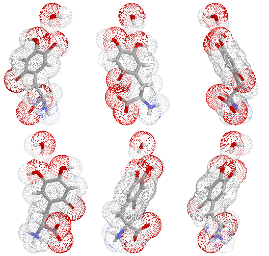
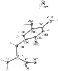
| F-DOPA
3,4-dihydroxy-6-fluoro-DL-phenylananine monohydrate 
|
 This structer was produced by RasMol, and stucked by GifBuilder (Yves Piguet, 1997) |
 ORTEP-III (Burnett, 1996) drawing |
Central delivery of 3,4-dihydroxy-L-phenylananine (L-DOPA) is a target in
therapeutic strategy for Parkinson's disease or in diagnoses for cognitive
disorder of brain. Radiofluorinated analogue of L-DOPA,
3,4-Dihydroxy-6-[18F]fluoro-L-phenylalanine ([18F]-L-DOPA) is used as a
tracer in the positron emission tomography (PET) studies of the dopamine
metabolism in the normal and pathological brain.1 It is assumed on the
basis of the biochemical and pharmacological similarity that the
distribution and metabolism of F-L-DOPA reflect those of L-DOPA.
We examine the crystal structure of [19F]-DL-DOPA as a stable
isotopic compound of [18F]-L-DOPA.
Paper: Crystal Structure of 3,4-Dihydroxy-6-fluoro-DL-phenylananine (F-DOPA) Monohydrate Used as a Positron Emission Tomography (PET) Imaging Ligand, Mitsunobu DOI, Masahiro SASAKI, Hisahiro KAWAI and Toshimasa ISHIDA Analytical Sciences (1998), 14, 1189-90. |
X-Ray Data Summary
| formula | C9 H10 F N O4 + H2O | ||
| weight | 233.20 | ||
| symmetry | monoclinic | ||
| space group | P21/c | ||
| Cell | Crystal | ||
| a (Ang) | 10.626(3) | description | needle |
| b (Ang) | 7.482(3) | colour | colorless |
| c (Ang) | 13.065(4) | size (mm) | 0.5x0.09x0.09 |
| alpha (deg) | 90.0 | Dx (g/ml) | 1.553 |
| beta (deg) | 106.23(2) | F(000) | 488 |
| gamma (deg) | 90.0 | mu(CuKa) | 1.206 |
| V (Ang^3) | 997.4(6) | ||
| Z | 4 | ||
| Refinement | Diffrn measurement | ||
| Flack | device type | Rigaku AFC5R/RU-200 | |
| parameters | 147 | decay | 0.7 % |
| restraints | 0 | index limit | -12-h-12,-8-k-0,-15-L-15 |
| R_factor_all | 0.0592 | theta (deg.) | 4.33 - 65.58 |
| R_factor_gt | 0.0453 | total reflections | 3239 include Friedel pairs |
| wR_factor_ref | 0.1321 | reflections(obs) | 1393 .gt. 2sigma(I) |
| wR_factor_gt | 0.1186 | Structure | |
| shift/su_max | lt.0.001 | solution | SHELXS-97 |
| shift/su_mean | lt.0.001 | refinement | SHELXL-97 |
Get PDB coordinates
Get CIF text
Get SHELXL INS file
 Back to the structure index
Date: Jun.1998