| C-Asn
Cytosine-(2-carboxyethyl)-1-yl-L-asparagine, monohydrate 
Date: Dec.1999 |
 This structer was produced by Tep2Rotater, and stucked by GifBuilder 0.5 (Yves Piguet, 1997) |
 ORTEP-III (Burnett, 1996) drawing | 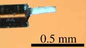 Photo of crystal used for data collection. |
Integration of the amino acid and the nucleic base has produced probes having outstanding properties to investigate the molecular recognition, and it is, for example, well known that the peptidic nucleic acids make thermostable complexes with DNA1. In our serial studies,2,3 the cooperation of the amino acids and the nucleic acids has been attempted to design the molecules which interact with the certain nucleic base, and the structures of the hybrid dipeptides including the cytosine base have been analyzed. In this paper, the structure of cytosine-hybrid dipeptide including L-asparagine (Fig.1) is reported. The title compound (C-Asn) was synthesized from 1-(2-carboxyethyl)-cytosine and asparagine methyl ester. After trials of the crystallization by changing the solvent conditions, only an aqueous methanol solution gave small crystals with the dimension of 0.05´0.05´0.20 mm3. These crystals insufficiently diffracted X-ray on the Rigaku RU200 generator. Intensity data was, therefore, measured on a synchrotron, SPring-8/BL24XU-A. The crystal and experimental data are listed in Table 1.
Paper: Crystal Structure of Cytosine Hybrid Dipeptide, Cytosine-(2-carboxyethyl)-1-yl-L-asparagine
Mitsunobu DOI, Akiko ASANO1, Toshimasa ISHIDA, Masahiro SASAKI, Masamichi NAKAI, Hiroshi HASEGAWA, Yoshio KATSUYA, Yoshihiro MEZAKI, Taizo TANIGUCHI and Akira TERASHIMA
Analytical Sciences, , in submission (2000)
X-Ray Data Summary
| formula | C11H15N5O5, H2O | ||
| weight | 315.30 | ||
| symmetry | orthorhombic | ||
| space group | P212121 | ||
| Cell | Crystal | ||
| a | 17.9558(16) Ang. | description | needle |
| b | 22.8603(19) Ang. | colour | Colorless |
| c | 4.9591(2) Ang. | size (mm) | 0.25x0.05x0.05 |
| alpha | 90.0 deg. | Dx (g/ml) | 1.029 |
| beta | 90.0 deg. | F(000) | 664 |
| gamma | 90.0 deg. | mu(CuKa) | 0.085 |
| volume | 2035.6(3) Ang^3 | wave length | 0.834 A |
| Z | 4 | temperature | 253(2) |
| Diffrn measurement | Refinementspace | ||
| device type | Rigaku RAXIS-4 | Flack | -0.2(19) |
| radiation source | SPring-8/BL24XU-A* | parameters | 210 |
| index limit | 0~h~22, 0~k~28, -5~L~5 | restraints | 0 |
| theta (deg.) | 3.41-31.41 | R_factor_gt | 0.0797 |
| total reflections | 3194 | wR_factor_gt | 0.2053 |
| reflections(obs) | 3086 .gt. 2sigma(I) | dela rho_max | 0.573 e A^3 |
| Structure | delta rho_min | -0.233 e A^3 | |
| solution | SHELXL-97 | shift/su_max | lt.0.001 |
| refinement | SHELXL-97 | Goodness of fit | 1.009 |
Get PDB coordinates
Back to the structure index
